

How researchers created “anthropobots” – What are their uses
Tiny living robots from human cells created by team of scientists. As CNN explains, they can move around in a petri dish (lab dish) and one day may be able to help heal wounds or damaged tissue, according to new research.
A team at Tufts University and Harvard University’s Wyss Institute have dubbed these creations “anthropobots.” The research builds on earlier work by some of the same scientists, who created the first living robots, or xenobots, from stem cells derived from embryos of the African clawed frog (Xenopus laevis).
“Some people thought that the characteristics of xenobots depended a lot on the fact that they are embryonic and amphibious,” said study author Michael Levin, the Vanevar Bush Professor of Biology at Tufts School of Arts & Sciences.
“I don’t think this has anything to do with being a fetus. This is not about being a frog. I think this is a much more general property of living things,” he clarified.
“We don’t realize all the abilities that our body’s cells have.” While living, the robots were not complete organisms because they did not have a complete life cycle, Levin said.
“It reminds us that these hard binaries we’ve worked with: Is it a robot, is it an animal, is it a machine? Such things do not serve us very well. We have to overcome it.”
How did they create them?
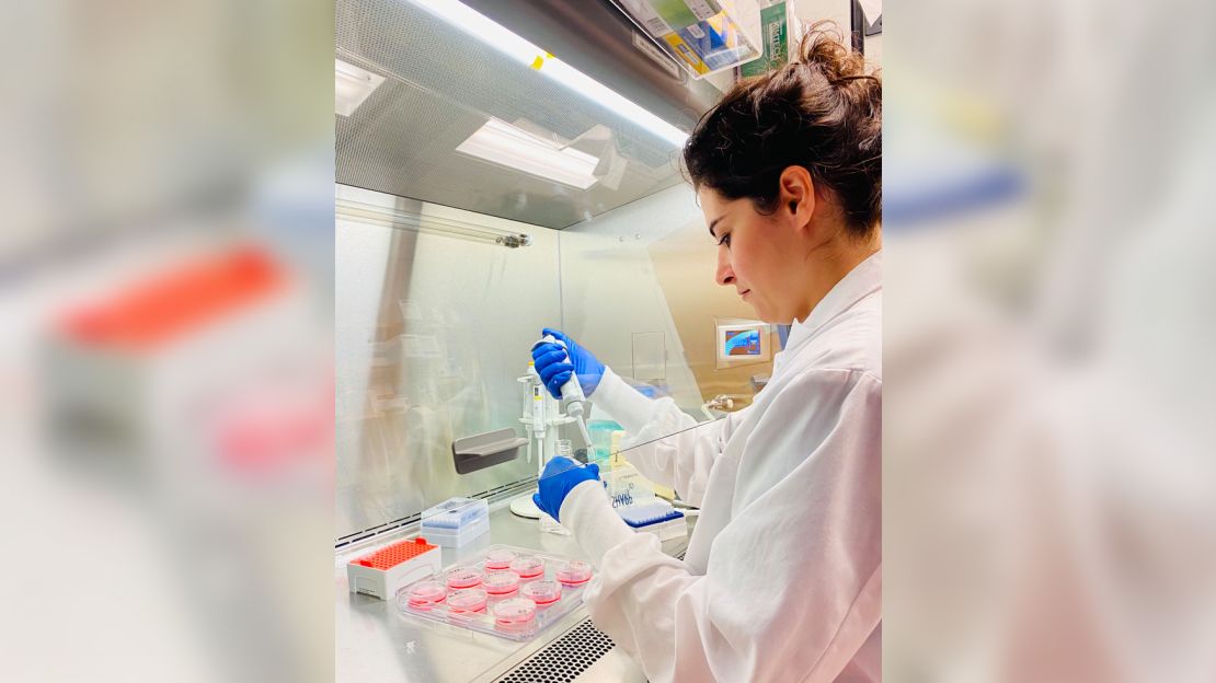
The scientists used adult human cells from the trachea, from anonymous donors of different ages and sexes. The researchers looked at this type of cell because they are relatively easy to access because of the work that has been done on Covid-19 and lung disease and, more importantly, because of a characteristic that scientists thought would make the cells able to move. , said study co-author Gizem Gumuskaya, a doctoral student at Tufts.
The cells of the trachea are covered with capillary projections called “cilia” that wave back and forth. They usually help cells in the trachea push out tiny particles that find their way into the air passages of the lungs. Previous studies have also shown that cells can form organelles – clumps of cells widely used for research.
The humanbots the team created were not identical, with different shapes and sizes.
Falk Tauber, team leader at the Freiburg Center for Interactive Materials and Bioinspired Technologies at the University of Freiburg in Germany, said the study provided a basis for future efforts to use bio-bots for different functions and to build them in different forms.
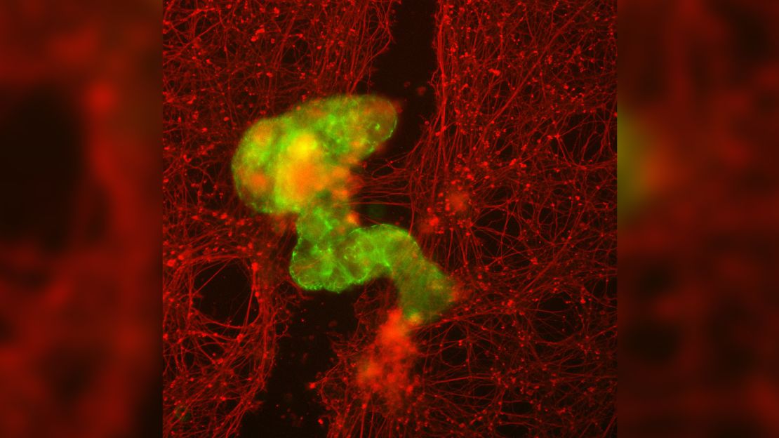
Tauber, who was not involved in the research, said the robots exhibited “unexpected behavior,” particularly when they moved into — and eventually closed — lacerations in human neurons.
He said the ability to create these structures from a patient’s own cells demonstrates diverse applications both in the lab and, eventually, in humans.
The research was published Thursday in the journal Advanced Science
Source :Skai
I have worked in the news industry for over 10 years. I have a vast amount of experience in covering health news. I am also an author at News Bulletin 247. I am highly experienced and knowledgeable in this field. I am a hard worker and always deliver quality work. I am a reliable source of information and always provide accurate information.


